How Much Drainage Is Normal for Chest Tube: A Practical Guide
Learn what drainage levels are considered normal after chest tube placement, how clinicians monitor drainage, warning signs to watch for, and when removal may be appropriate.

Normal chest tube drainage is not a fixed volume. In most cases, drainage is higher right after insertion and gradually declines over the first day or two. A wide range—from small serous amounts to several hundred milliliters per day—can be expected early on, depending on injury, surgery, and suction use. The key is a downward trend and no new air leaks.
How much drainage is normal for chest tube
In clinical practice, the exact volume of drainage after chest tube placement depends on the underlying problem and surgical factors. Normal drainage is not a fixed number; clinicians look for a downward trend over time and for absence of new air leaks. In the initial hours after insertion, drainage can be more noticeable due to blood, fluid, or air evacuation, but it should begin to wane as healing progresses. The question of how much drainage is normal for chest tube depends on the indication—pneumothorax, hemothorax, or postoperative drainage—and on whether suction is used. Clinicians also consider the patient’s overall trajectory, imaging results, and clinical stability. Drainage amount is one piece of the puzzle; color, clarity, and the presence or absence of air leaks are equally important signals of progress.
Factors that influence drainage volumes
Drainage volume is influenced by several variables: the cause of chest tube placement (trauma, surgery, infection), the size and type of chest tube, whether suction is applied, and how long the tube has been in place. Blood-rich specimens will produce higher initial volumes, while serous or serosanguinous fluid tends to decrease more quickly. Activity level, lung re-expansion, and the patient’s ability to heal also play roles. The timing of removal is closely linked to both the drainage amount and the radiographic picture. Clinicians monitor trends rather than isolated numbers to guide decisions, and they adjust suction or drainage strategy based on patient progress.
Monitoring and documentation: how clinicians track drainage
Care teams document the drainage volume at regular intervals, color and consistency, and the presence of air leaks. Drainage can be logged hourly, especially in the first 24 hours, and then every 4-6 hours as patient status stabilizes. Imaging studies, such as chest radiographs, are used alongside drainage data to confirm lung re-expansion and absence of residual collections. Consistent records help detect deviations from expected trends early, which is critical for preventing complications. Nurses and physicians work together to interpret the total clinical picture, not just the raw numbers.
Red flags signaling the need for reassessment
Seek prompt medical attention if you notice a sudden increase in drainage, a change to brighter red color, or large clots in the collection chamber. A persistent air leak beyond 24-48 hours, fever, chest pain, or shortness of breath can indicate complications or incomplete lung re-expansion. These signs warrant immediate communication with the care team and possible adjustment of suction, imaging, or consideration of alternative interventions. Any new symptoms in conjunction with drainage changes should be treated as a warning signal.
Patient experiences during chest tube drainage
Patients may experience pain or discomfort around the insertion site, breathlessness, and fatigue. Effective analgesia, careful positioning, and breathing exercises help manage symptoms while routine drainage monitoring continues. Family members should understand that some variation in drainage and minor fluctuations are normal in the early days. Clear communication with nurses and physicians is essential for reassurance and timely reporting of concerns.
Removal decisions and timelines
Chest tube removal is based on clinical and radiographic criteria: drainage should be minimal or absent for a defined period (often 24 hours) and lung re-expansion should be confirmed. If an air leak persists or imaging shows unresolved issues, removal may be delayed. Decisions are tailored to the patient, procedure, and stability, with a structured plan documented in the medical record. In some cases, suction may be weaned first, then the chest tube is removed when safe and indicated.
Practical tips to participate in care
Ask questions about what the numbers mean and how your treatment will progress. Keep a simple diary of daily drainage measurements, color changes, and symptoms to discuss at rounds. If you’re supporting a loved one, help them stay comfortable, communicate concerns promptly, and follow instructions on activity and coughing techniques to support lung expansion. Understanding the drainage trajectory helps patients stay engaged and reduces anxiety during recovery.
Overview of drainage expectations by phase
| Aspect | Typical Range / Guidance | Notes |
|---|---|---|
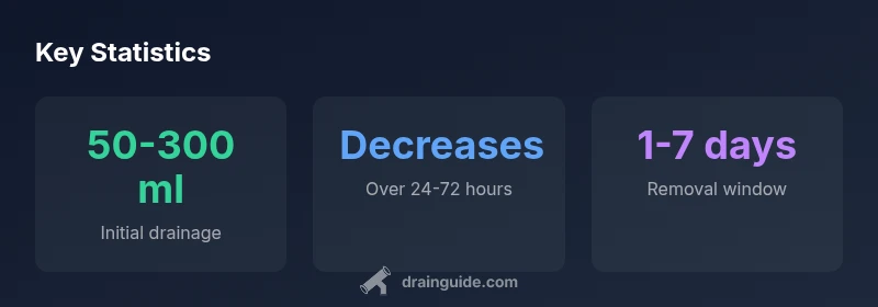
| Initial drainage (first 24 hours) | 50-300 ml | Higher with extensive thoracic injury or surgery |
| Drainage trend (48 hours) | Decreases steadily | Supports removal when minimal for 24 hours |
| Air leak duration | Hours to days | Persistent leak requires evaluation |
| Removal readiness | Typically after minimal drainage for 24 hours | Guided by imaging and clinical status |
Got Questions?
What counts as normal drainage after chest tube placement?
Drainage varies by procedure and patient, but most people see higher volumes initially that gradually decline. A downward trend with stable or improving imaging is a positive sign. If there are sudden changes or new symptoms, notify the care team.
Drainage varies, but a downward trend and stable imaging are good signs. Tell your clinician if you notice sudden changes.
How long does drainage typically last?
Drainage typically tapers over 1-3 days, with removal considered once drainage is minimal for about a day and imaging confirms lung expansion. Individual recovery can vary.
Drainage usually tapers in a day or two, but it varies by patient.
What should I do if drainage seems to stay high after 2 days?
Persistent high drainage requires clinical evaluation. The care team may adjust suction, order imaging, or reassess for complications. Do not delay reporting these signs.
If drainage stays high after a couple days, tell your doctor.
Can suction affect drainage volume?
Yes. Suction and gravity drainage influence the volume collected. Any changes should be coordinated with the care team to avoid misinterpretation.
Suction can change how much drains; alert your team.
Is chest tube drainage painful?
Pain is common after chest tube placement. Pain control is a priority, and analgesia should be adjusted to keep you comfortable while monitoring drainage.
Yes, it can be painful; your team will manage it.
When is removal of the chest tube considered?
Removal is usually considered after minimal drainage for 24 hours and satisfactory radiographic results. Clinical status guides the final decision.
Removal is considered once drainage is minimal and imaging is good.
“Drainage after chest tube placement is highly patient-specific; clinicians look for a consistent downward trend and no new air leaks when considering removal.”
The Essentials
- Drainage should trend downward after chest tube placement.
- Monitor volume, color, and air leaks daily.
- Removal is guided by minimal drainage and imaging findings.
- Report sudden changes to your care team.